While a normal aortic valve has three leaflets, a bicuspid aortic valve (BAV) is a congenital anomaly where the valve has only two leaflets. It results from an embryonic development defect with a strong genetic component, often linked to the NOTCH1 gene.
BAV is the most common congenital heart defect, affecting about 2% of the population, and is more prevalent in males. At birth, the bicuspid valve typically functions normally and remains asymptomatic for years. About half of patients also have some degree of ascending aortic dilation.
Consequences of a Bicuspid Aortic Valve
- Over time, the bicuspid valve deteriorates, becoming fibrotic or even calcified, leading to narrowing (aortic stenosis) and/or leakage (aortic regurgitation).
- It rarely causes issues during childhood, making it a source of concern for parents, but often asymptomatic for the child.
- Severe valve dysfunction usually appears later in life, typically after the age of 40.
The bicuspid valve is constantly subjected to stress and friction with each heartbeat, which may lead to progressive fibrosis, deformation, and calcification. This causes the valve to open less and/or fail to close completely. As a result, the left ventricle becomes chronically overloaded, leading to hypertrophy and/or dilation.
Severe valve dysfunction generally arises in adulthood, but in rare cases, it can manifest in childhood or adolescence, most frequently as aortic stenosis, though aortic regurgitation can also occur. Extreme cases are evident at birth, especially with unicuspid valves—effectively a membrane with a small orifice.
Patients with BAV and ascending aortic dilation also face risks from the aortic abnormality. Aortic dilation may progress, leading to aneurysm, wall thrombosis, dissection, or rupture, which can be fatal regardless of the valve’s condition.
Diagnosis of Bicuspid Aortic Valve
- Diagnosis often begins when a heart murmur is detected during auscultation.
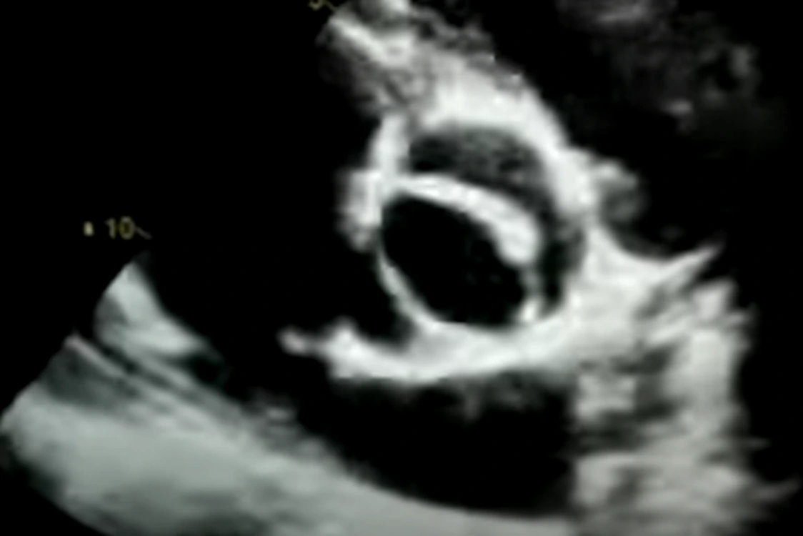
- Echocardiography is the gold standard for confirming BAV and is recommended for all children with murmurs.
- Usually, no other tests are needed; monitoring involves periodic echocardiograms.
- Frequent imaging is unnecessary unless the valve shows significant deterioration.
- In advanced cases, echocardiograms are repeated every 6–12 months.
- Echocardiography also detects ascending aorta dilation, which is commonly associated with BAV.
Bicuspid valves do not always have the same shape: sometimes there are two real cusps, other times three cusps with one fused commissure. The orientation of cusps or fused commissures also varies, leading to five recognized BAV types.
There is a strong correlation between BAV morphology and the severity of valve dysfunction or aortic dilation. Blood flow direction through the valve varies with each type, possibly explaining these differences. A genetic link is also suspected. A complete BAV evaluation is advised to predict the disease’s progression and aortic impact.
Transthoracic echocardiography (TTE) is the primary imaging tool for BAV, but it has limitations in resolution and in assessing the entire aorta. If imaging is difficult—especially with aortic dilation—a transesophageal echocardiogram (TEE) may be used for better visualization of both the valve and thoracic aorta.
Other advanced imaging techniques such as CT angiography and cardiac MRI provide excellent images of the aortic valve and thoracic aorta throughout its full length.
Treatment of Bicuspid Aortic Valve
Patients with BAV should maintain a healthy lifestyle to slow the progression of valve deterioration. A diet low in salt and saturated fat, and rich in fruits, vegetables, and fish, is recommended. Cholesterol and blood pressure should be well-controlled, as hypertension accelerates aortic dilation.
Treatment depends on the type and severity of valve damage. In childhood cases of severe aortic stenosis with non-calcified cusps, surgery is not always required. Instead, a balloon valvuloplasty via cardiac catheterization may be performed. This minimally invasive procedure avoids general anesthesia and open surgery.
If the valve is severely calcified or fails to close properly (aortic regurgitation), and dysfunction is advanced, surgical valve replacement is necessary. The diseased valve is removed and replaced with a prosthetic valve, either biological (from human/animal tissue) or mechanical (made from synthetic materials).
Surgical decisions also depend on the extent of aortic dilation. Surgery is typically recommended if the ascending aorta measures 55 mm or more—though many experts opt for surgery at 50 mm. If aortic valve surgery is already indicated, ascending aorta replacement is often performed when the diameter is 45 mm or more.
Prognosis
The prognosis for patients with bicuspid aortic valve is usually excellent, with normal life expectancy—provided severe deterioration does not occur, or surgical/valvuloplasty outcomes are favorable.
This article is for educational purposes only and does not replace medical consultation.